The following is an introduction to the examination equipment used at the Hoshii Ophthalmology Department.
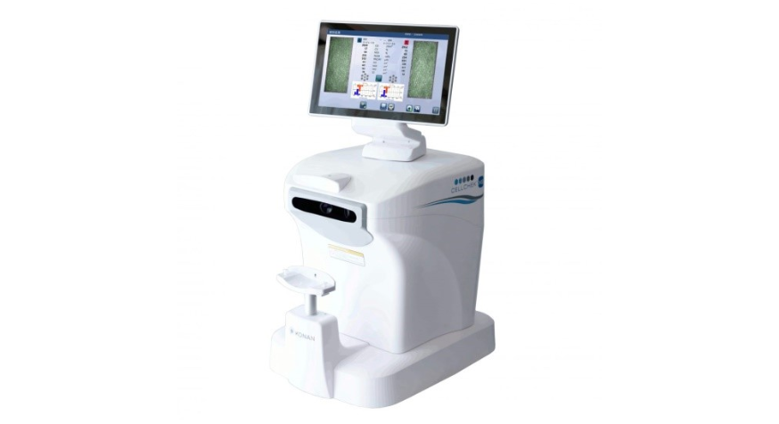
Specular Microscope
Automated analysis of corneal endothelial cells using the highly reliable 'Center Method'. Accurately diagnose corneal endothelial effects after contact lens or surgery in a small amount of time.
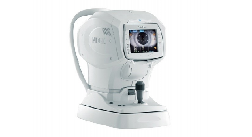
Autorefucherat/tonometer TONOREF® II
Highly accurate data acquisition of refractive status for farsightedness, myopia, and astigmatism is possible through 3D auto-tracking and auto-measurement. At the same time, intraocular pressure can be measured by patient-friendly air discharge.
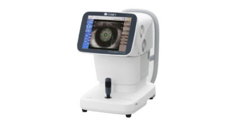
Optical Ocular Axis Length Measuring Device OA-2000
Many areas necessary for cataract surgery, such as ocular axis length (eye length), corneal roundness, anterior chamber depth, and lens thickness, are measured at once without touching the eye.
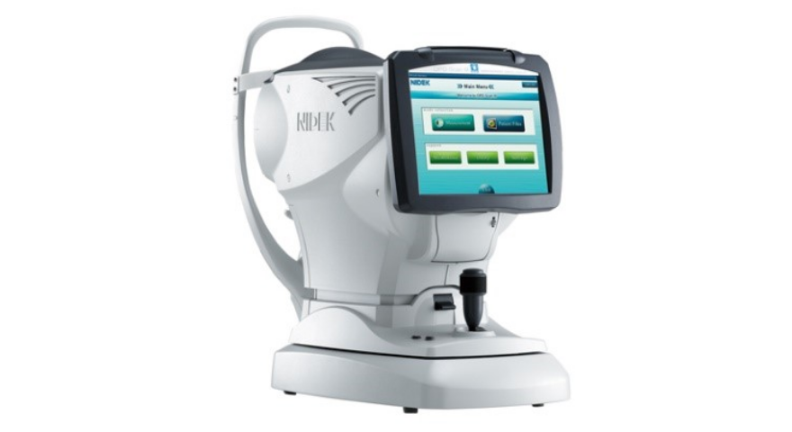
Corneal shape/refractive power analyzer OPD-Scan® III
Measures corneal shape (topo) and refractive power (ref) distribution for farsightedness, nearsightedness, and astigmatism. A vision simulation function is available to simulate the patient's vision from multiple angles.
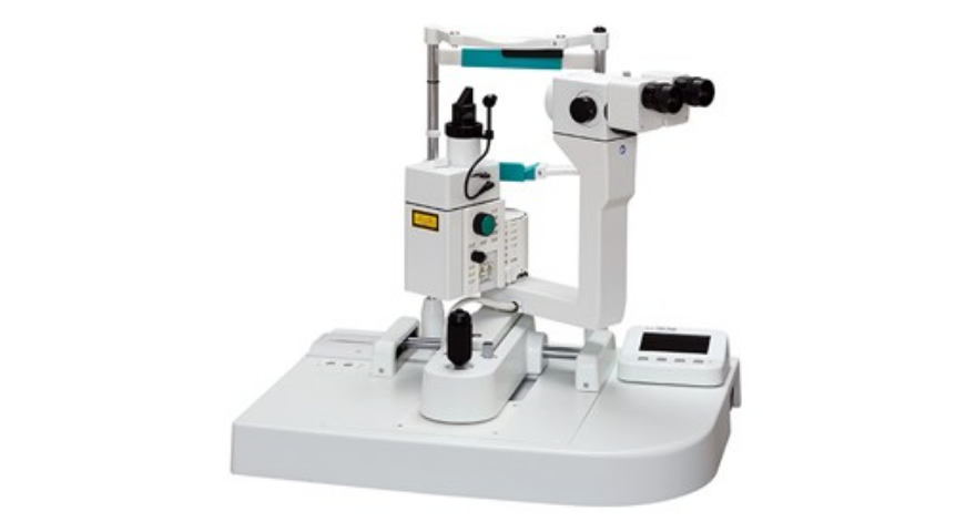
Anterior chamber protein analyzer Laser Flare Meter®.
Kowa FM-700
Non-contact, non-invasive quantitative measurement of protein concentration in the space called the anterior chamber of the eye.
Effective for early detection of anterior chamber inflammatory disease, evaluation of postoperative inflammation, and determination of treatment efficacy.
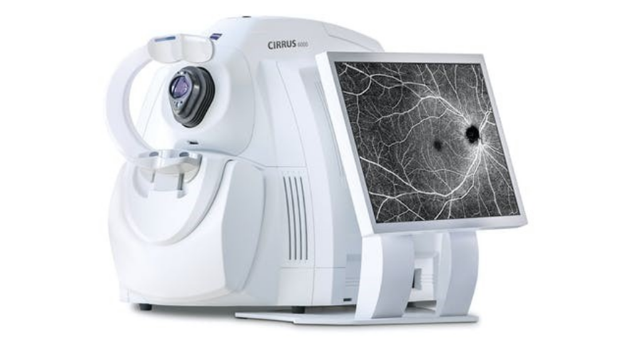
Optical Coherence Tomography Cirrus HD-OCT 6000
High-resolution SD-OCT, which provides a detailed view of the vitreous interface and retinal surface layers, and OCT Angiography (OCTA) scan images, which provide a more detailed view of retinal microvasculature.
Useful for diagnosis and treatment of age-related macular degeneration, diabetic retinopathy, and glaucoma.
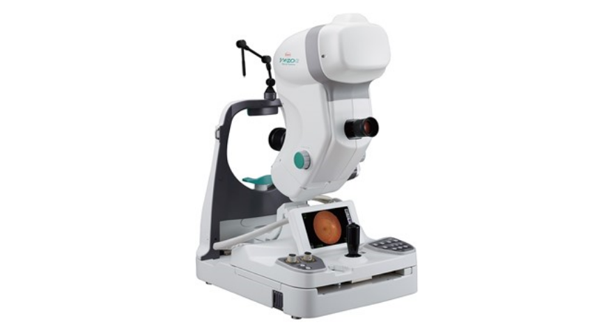
Mydriatic and non-mydriatic fundus camera Kowa VX-20α
Capable of taking fundus color photographs, contrast-enhanced fluorescent fundus photographs, and autofluorescent photographs. Retinal diseases are clearly photographed.
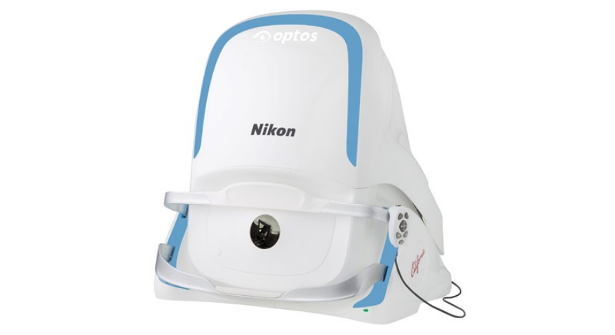
Ultra wide angle scanning laser ophthalmoscope Optos California
With an angle of view of 200 degrees, the camera captures approximately 80% of the fundus without mydriasis or contact. Capable of capturing a wider area than a conventional fundus camera in a single shot.
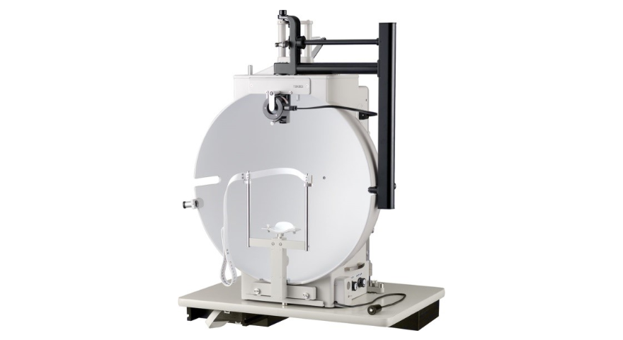
Goldmann Visual Field Meter (Dynamic Visual Field Testing)
Capable of measuring the sensitivity of the entire field of view.
Useful for detecting various visual field abnormalities as well as glaucoma.
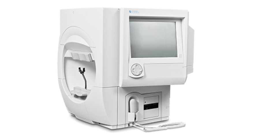
Humphrey visual field meter (static visual field testing)
Capable of measuring the sensitivity of the field of view within the center 30 degrees.
Integration with the electronic medical record has made it possible to clearly monitor the progression of glaucoma vision.
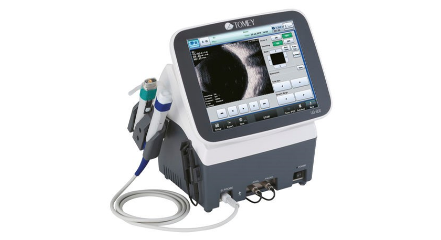
Ultrasound Imaging Diagnostic Ocular Axis Length and Corneal Thickness Measurement System UD-800
High frequency components (harmonic components) are extracted for high-resolution intraocular imaging.
Extracts detailed structures such as vitreous hemorrhages, opacities, retinal detachments, and intraocular tumors.
The following is an introduction of surgical instruments used at Hoshii Ophthalmology.
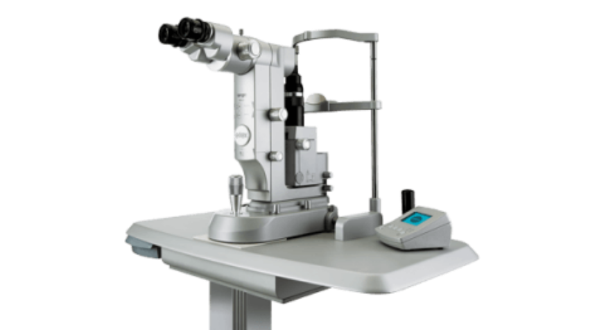
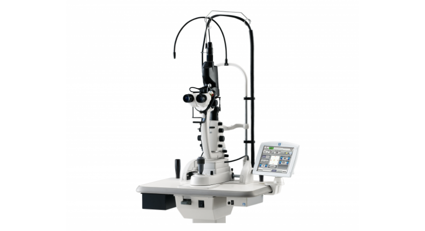
YAG (YAG) laser surgery equipment
Efficient low power level and low number of irradiations allow for lens saccotomy and iridotomy.
Useful in the treatment of late-onset cataracts and acute glaucoma attacks.
Multi-color scan laser photocoagulator MC-500 Vixi™.
Stable laser output enables short-time high-power pattern irradiation and 22 types of scan patterns for selection of patterns suitable for complex retinal diseases.
Useful in the treatment of diabetic retinopathy and retinal rupture.
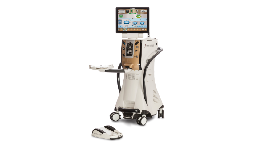
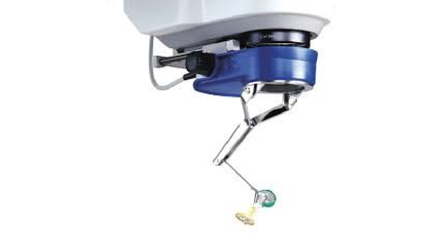
Cataract Surgery Device CENTURION® Vision System
Designed for safety, stability, and controllability, it offers a new dimension in cataract surgery.
High anterior chamber stability and consistent intraocular pressure are maintained for minimally invasive and highly efficient surgery.
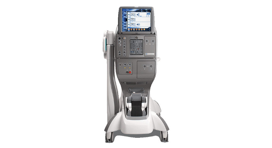
Vitrectomy System Constellation® Vision System
Real-time perfusion adjustment with the IOP control function allows for new intraocular pressure stability and a smoother, more efficient vitrectomy setup.
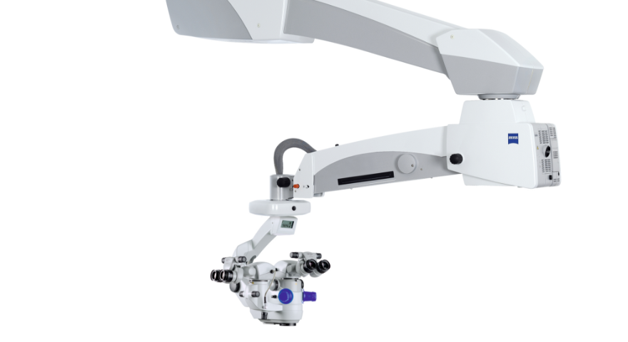
Operating Microscope OPMI Lumera 700
The microscope enables more precise and safer ophthalmic surgery by allowing the surgeon to observe the surgical site more clearly and accurately than with a conventional microscope, and to check detailed movements and eye conditions during surgery with a clear field of view.
Resight (700)
It provides a clear image of the state of the retina. Integration with the Lumera 700 allows for easy focusing through the microscope.
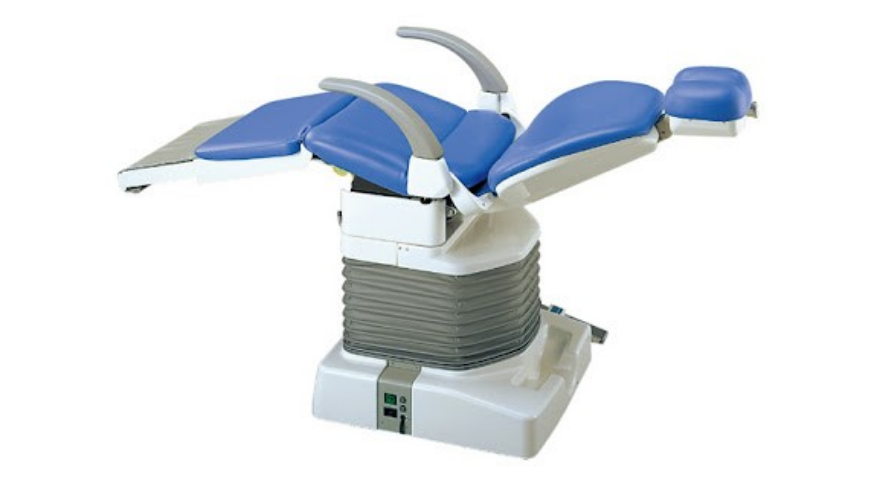
Ophthalmic operating table TAKARA BELMONT MEPRO
The chair-type operating table helps patients move from the introduction to the surgical position.
Allows surgery to be performed in a more stable position.